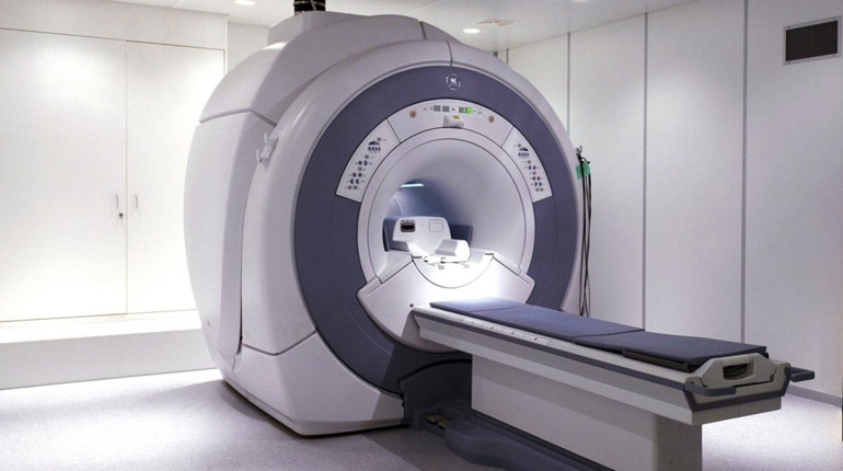
MRI is a modern method of examining the brain

MRI of the brain is one of the modern non-contact methods of brain research, which uses a magnetic field and radio waves that transmit a signal to a computer and allow you to assess the state of the brain. It is important to know that MRI of the brain is used to examine both soft tissues and blood vessels for damage or injury, such as a stroke.

What is the best time to have an MRI?

MRI of the brain is performed to detect or confirm a wide range of diseases. During the MRI examination, the doctor sees a detailed image of the brain on the screen, assesses its condition and can detect pathological diseases.

Sometimes, MRI diagnosis may be required to confirm or refute the diagnosis.

If:

– Acute or constant headaches.

– There is a constant or periodic noise in the ears

– there is weakness and numbness in the extremities;

– there is a deterioration of memory;

– fainting occurs periodically;

– The person is confused;

– There was a craniocerebral injury.

– you need to find out the cause of seizures.

Contrast-enhanced MRI of the brain

Contrast is used to better see the brain. It is introduced into the body. The dye can be used to diagnose tumors and other diseases, as well as their structure and contours.

Contraindications to Use

MRI of the brain can be the most safest, but it is not recommended for everyone.

Also, it is worth quitting MRIs when:

– pregnancy;

– claustrophobia;

– The presence of cochlear implant;

– decompensated heart failure;

– the presence of tattoos created on a metal basis;

Installed crowns and braces

What is an MRI exam?

The preparation is the first step in the examination. It is important to take out all metal objects from the phone and then remove it.

After the patient is placed on the table, and a device is fixed on the head, which will send and receive radio waves. MRI is performed for 30-60 minutes, depending on the department and the presence of contrast in the body.

The doctor takes many layers-by-layer photographs of the brain and then makes a decision.

More information about MRI of the brain just go to this popular resource: click to read more

How to cure a hernia without surgery

The diagnosis of an intervertebral disc herniation sounds like a sentence to some people. Instantly, the picture of surgical and operating instruments is drawn in the head. In fact, everything is not so scary.

What is an intervertebral hernia and what are its causes

Lumbar spine hernia, and other localizations, are protrusions from the disc nucleus. They result from partial or complete ruptures of the fibrous outer shell. Hernia, provoked by excessive physical activity, is dangerous risk of restriction of the nerve roots of the spinal cord.

People at risk of developing an intervertebral hernia are:

* elderly;

* overweight;

* have poor heredity.

Work can lead to herniated vertebrae. The problem often arises in professional weightlifters, loaders, truck drivers. A lack of exercise or sedentary lifestyle may lead to atrophy of the back muscles.

People with healthy habits and who are active in sports rarely need to see a neurologist for such issues.

Symptoms of lumbar hernia

Lumbar hernia is manifested by back pain of varying intensity and other symptoms.

1. Numbness of soft tissues on the outer surface of the thigh, shin and big toe with a tingling sensation and tingling.

2. Numbness or sharp pain in the lower back and buttocks join the numbness.

3. Mobility of the vertebrae in lower parts is restricted.

4. Painful shots can be taken even if you are lifting very small weights off the ground in your waist.

5. The patient experiences general weakness and fatigue quickly. Sensitivity decreases in the groin area.

Intervertebral hernia in the cervical region:

Hernia in the cervical spine manifests itself in the early stages.

1. Even if the patient is not turning, severe pain can be felt. Therefore, it is a good idea to return your entire torso.

2. The pain radiates from the wrist to the shoulder, where it is almost completely gone.

3. Numbness occurs when you squeeze the brush.

4. The patient complains of headaches, flies in the eyes, tinnitus, loss of orientation in space.

The main symptoms may be accompanied by a hypertensive crisis.

It is not necessary to have all the signs at once, and a visual inspection will not give anything. X-rays, MRI and CT are used to diagnose the condition. A CT will clearly show a protrusion in between the vertebrae. In some cases, electroneuromyography may be required, a diagnostic procedure that can assess the degree of nerve root compression.

Is it possible to treat intervertebral hernia with out surgery?

A complete exam is required before the doctor can determine if it is possible to treat intervertebral hernia with surgery or if it is better to go to surgery. In advanced cases, surgery is the only way to relieve pain, restore mobility and efficiency.

The Medical Diagnostic Center “Diagnostics”, Kiev, for intervertebral hemia treatment without surgery uses the best techniques. The clinic’s doctors also prescribe physiotherapy and massage to patients. They also create individual therapeutic gymnastics complexes. In other words, in each case the therapeutic program is developed individually and guarantees positive dynamics.

For additional information about gryzha diska explore this useful net page

MRI is a modern method of examining the brain

The modern non-contact method of brain research is MRI of the Brain. It uses radio waves and a magnetic field to transmit a signal to the computer that allows you to assess the brain’s state. It is important that MRI of brain can be used to assess the health and condition of both soft tissues as well as blood vessels.

What is the best time to have an MRI?

MRI of the brain is performed to detect or confirm a wide range of diseases. During the MRI examination, the doctor sees a detailed image of the brain on the screen, assesses its condition and can detect pathological diseases.

Sometimes, MRI diagnosis may be required to confirm or refute the diagnosis.

MRI should be performed if:

– disturbed by constant or acute headaches;

– there is a periodic or constant noise in the ears;

– There is weakness and numbness at the extremities.

– There is a decline in memory.

– fainting occurs periodically;

– The person is confused;

– there was a craniocerebral trauma;

– you need to find out the cause of seizures.

Contrast-enhanced MRI (MRI) of the brain

In order to see the brain more clearly, a contrast is used, which is introduced into the human body. The dye can be used to diagnose tumors and other diseases, as well as their structure and contours.

Contraindications to Use

MRI of the brain can be the most safest, but it is not recommended for everyone.

It is also worth giving up MRI when:

– pregnancy;

– claustrophobia;

– The presence of cochlear implant;

– Decompensated Heart Failure

– The presence of tattoos that are made from metal;

– installed crowns or braces.

What is an MRI exam?

The preparation is the first step in the examination. It is important to take out all metal objects from the phone and then remove it.

After the patient is placed on the table, and a device is fixed on the head, which will send and receive radio waves. MRI is performed for 30-60 minutes, depending on the department and the presence of contrast in the body.

The doctor takes many layers-by-layer photographs of the brain and then makes a decision.

For more info about MRI of the brain browse this popular net page

What does an MRI of the spine show?

MRI with the spine is important to help make a precise diagnosis and prescribe the best treatment option. Laptop computer is one of the most informative, but requires some preparation and proper interpretation from the results.

INDICATIONS

MRI from the spine is prescribed the if you have a suspicion of the pathology in the ridge. The analysis is desirable for trauma, various developmental abnormalities, inflammatory diseases, degenerative processes, malignant formations, metastases.

The process is needed:

– in the event of severe lumbar pain;
– shooting or aching pains with recoil within the thigh, calf, groin or buttocks;
– incontinence of feces and urine;
– pinching and loss in mobility.

Magnetic resonance imaging is prescribed following your patient has been examined by way of a neurologist.

Exactly what does MRI SHOWS?
A radiologist or even a doctor of functional diagnostics deals with decoding of MRI images of the spine. Three-dimensional cards are compared with pictures of a healthy person, and possible pathological changes are identified. Such as: hernia, osteochondrosis, etc. The analysis will help determine the stage of growth and development of the disease, along with choose the best treatment options. Around the cards, you can clearly understand the soft tissues and bones – the bones are painted in the dark color, as well as the vertebrae is within light colors.

What’s DISPLAYED From the IMAGES?

Many people are enthusiastic about exactly what the MRI of the spine shows. The method will show the following results:

– just how much possible damage to the spine, as well as the existing pathologies. It is possible to identify them in early stages;
– see neoplasms and possible inflammation in soft tissues;
– to look for the nature and extent from the injury;
– to recognize a hernia, tomography shows the protrusion with the muscles and longitudinal ligaments.

How can an MRI WORK?

For magnetic resonance imaging, the individual is placed within a special apparatus, where the area of ??our bodies under investigation is scanned by using a magnetic field. Info is saved, printed, visualized, after which receives for analysis by way of a doctor. The process doesn’t cause discomfort, but throughout the MRI you’ll want to lie still for your image to be of excellent quality. Normally the research takes most an hour.

PREPARATION

You should lift off all metal objects: rings, earrings, watches, etc. Mobile phones ought to be left outside of the premises. Several hours prior to the diagnosis, it’s not necassary to take food, medications, or drink liquids. It is recommended wear loose-fitting clothing it doesn’t hinder movement. The examination is utterly painless, and you may get rid of unpleasant sounds in the operation from the tomograph with the aid of earplugs.

Contraindications

Absolute contraindications add the presence of electronic implanted medical devices, ferromagnetic heart valves, a good massive ferromagnetic medical structures in the body.

Relative contraindications include pregnancy, the use of metal structures within the skeleton, dentures, prosthetic heart valves, insulin pumps and nerve stimulants.

To learn more about MRI of the spine browse our web page: click now

How to cure a hernia without surgery

The diagnosis of an intervertebral disc herniation sounds like a sentence to some people. Instantly, the picture of surgical and operating instruments is drawn in the head. In fact, everything is not so scary.

What is an intervertebral hysteria? And what are its causes?

Lumbar spine hernia, and other localizations, are protrusions from the disc nucleus. They result from partial or complete ruptures of the fibrous outer shell. Hernia, provoked by excessive physical activity, is dangerous risk of restriction of the nerve roots of the spinal cord.

People at risk of developing an intervertebral hernia are:

* elderly;

* overweight;

* have poor heredity.

Herniated vertebrae can be the result of work. Professional weightlifters, loaders, and truck drivers are often affected by this problem. The cause of the pathology may be atrophy of the back muscles due to sedentary lifestyle, lack of exercise.

People without bad habits, who play sports and lead a healthy lifestyle, rarely consult a neurologist with such problems.

Lumbar hernia symptoms

Lumbar hernia can manifest as back pain with varying intensity or other symptoms.

1. Tingling sensations and tingling caused by numbness in the soft tissues of the outer surface of the thigh, big toe and shin.

2. The numbness is accompanied by numbness and sharp pains in the buttocks and lower back.

3. Mobility of the vertebrae in lower parts is restricted.

4. Painful shots can be taken even if you are lifting very small weights off the ground in your waist.

5. The patient experiences general weakness and fatigue quickly. Sensitivity decreases in the groin area.

Symptoms of intervertebral hernia of the cervical region

In the beginning stages, hernia can occur in the cervical spine.

1. Even if the patient is not turning, severe pain can be felt. Therefore, it is a good idea to return your entire torso.

2. The pain radiates from the wrist to the shoulder, where it is almost completely gone.

3. Numbness occurs when you squeeze the brush.

4. The patient complains of headaches, flies in the eyes, tinnitus, loss of orientation in space.

Hypertension may accompany the main symptoms.

You don’t have to see all signs at once. A visual inspection won’t give any information. Diagnosis is performed by X-ray, MRI and CT. An X-ray will clearly show a protrusion between the vertebrae. In some cases, electroneuromyography may be required, a diagnostic procedure that can assess the degree of nerve root compression.

Is it possible to treat intervertebral hernia with out surgery?

A complete exam is required before the doctor can determine if it is possible to treat intervertebral hernia with surgery or if it is better to go to surgery. In advanced cases, surgery is the only way to relieve pain, restore mobility and efficiency.

The Medical Diagnostic Center “Diagnostics”, Kiev, for intervertebral hemia treatment without surgery uses the best techniques. Along with drug therapy, the clinic’s specialists prescribe massage and physiotherapy to patients, and make individual complexes of therapeutic gymnastics. In other words, in each case the therapeutic program is developed individually and guarantees positive dynamics.

For more info about gryzha poyasnichnogo otdela have a look at our new web portal

What does an MRI of the spine show?

MRI of the spine is necessary to make an accurate diagnosis and prescribe the best treatment option. The survey is among the most informative, but requires some preparation and proper interpretation from the results.

INDICATIONS

MRI with the spine is prescribed in almost all cases if you have a suspicion of the pathology from the ridge. The analysis is desirable for trauma, various developmental abnormalities, inflammatory diseases, degenerative processes, malignant formations, metastases.

The procedure is needed:

– in case of severe back pain;
– shooting or aching pains with recoil from the thigh, calf, groin or buttocks;
– incontinence of feces and urine;
– pinching and decrease of mobility.

Magnetic resonance imaging is prescribed as soon as the patient has become examined by the neurologist.

Precisely what does MRI SHOWS?
A radiologist or a doctor of functional diagnostics works with decoding of MRI pictures of the spine. Three-dimensional cards are in contrast to pictures of a normal person, after which possible pathological changes are identified. Included in this are: hernia, osteochondrosis, etc. The learning can help determine activity is of progression of the disease, along with select the right treatment options. For the cards, you are able to clearly start to see the soft tissues and bones – the bones are painted in a dark color, along with the spine is light colors.

Precisely what is DISPLAYED Within the IMAGES?

Many people are enthusiastic about just what the MRI with the spine shows. The task shows these results:

– the quality of possible harm to the spine, and also the existing pathologies. It is possible to identify them in the early stages;
– see neoplasms and possible inflammation in soft tissues;
– to discover the nature and extent from the injury;
– to realize a hernia, tomography can have the protrusion of the muscles and longitudinal ligaments.

How can an MRI WORK?

For magnetic resonance imaging, the person is positioned inside a special apparatus, in which the area of ??our bodies under investigation is scanned using a magnetic field. Info is saved, printed, visualized, and after that receives for analysis with a doctor. The method won’t cause discomfort, but during the MRI you should lie still to the image to be of proper quality. Usually research takes about half 1 hour.

PREPARATION

You should remove all metal objects: rings, earrings, watches, etc. Mobile phones also need to be left beyond your premises. A few hours ahead of the diagnosis, you shouldn’t take food, medications, or drink liquids. It is recommended wear loose-fitting clothing that doesn’t hinder movement. The examination is utterly painless, and you will get rid of unpleasant sounds in the operation in the tomograph by making use of earplugs.

Contraindications

Absolute contraindications are the existence of electronic implanted medical devices, ferromagnetic heart valves, a good massive ferromagnetic medical structures by the body processes.

Relative contraindications include pregnancy, a good metal structures from the skeleton, dentures, prosthetic heart valves, insulin pumps and nerve stimulants.

For more details about MRT pozvonochnika go to our new web page: look at more info

MRI is a modern method of examining the brain

MRI of the brain is one of the modern non-contact methods of brain research, which uses a magnetic field and radio waves that transmit a signal to a computer and allow you to assess the state of the brain. It is important that MRI of brain can be used to assess the health and condition of both soft tissues as well as blood vessels.

What is the best time to have an MRI?

MRI of the brain is performed to detect or confirm a wide range of diseases. During an MRI examination, the doctor can see a detailed image of your brain and assess its condition.

Sometimes, MRI diagnosis may be required to confirm or refute the diagnosis.

MRI should be performed if:

– Acute or constant headaches.

– There is a constant or periodic noise in the ears

– there is weakness and numbness in the extremities;

– There is a decline in memory.

Occasionally, fainting can occur

– the person is disoriented;

– there was a craniocerebral trauma;

– you need to find out the cause of seizures.

Contrast-enhanced MRI (MRI) of the brain

Contrast is used to better see the brain. It is introduced into the body. Thanks to the introduction of the dye, tumors and other pathological formations can be diagnosed, and their size, structure and contours can be studied in more detail.

Contraindications to use

MRI of the brain is the safest procedure, but some people do not do it: if they have a pacemaker, a hemostatic clip in the brain or metal implants.

It is also worth giving up MRI when:

– pregnancy;

– claustrophobia;

– the presence of cochlear implants;

– decompensated heart failure;

– The presence of tattoos that are made from metal;

Installed crowns and braces

What is an MRI exam?

The examination begins with the preparation: it is necessary to remove all metal objects and remove the phone.

After the patient is placed on the table, and a device is fixed on the head, which will send and receive radio waves. Depending on the area and whether there is contrast, MRI can take between 30-60 minutes.

The doctor takes many layers-by-layer photographs of the brain and then makes a decision.

To learn more about MRI of the brain take a look at this useful net page: read

How to cure a hernia without surgery

The diagnosis of an intervertebral disc herniation sounds like a sentence to some people. Instantly, the picture of surgical and operating instruments is drawn in the head. In fact, everything is not so scary.

What is an intervertebral hernia and what are its causes

Lumbar spine hernia, and other localizations, are protrusions from the disc nucleus. They result from partial or complete ruptures of the fibrous outer shell. Hernia, provoked by excessive physical activity, is dangerous risk of restriction of the nerve roots of the spinal cord.

People at risk of developing an intervertebral hernia are:

* elderly;

* overweight;

* Have poor genetics.

Herniated vertebrae can be the result of work. The problem often arises in professional weightlifters, loaders, truck drivers. The cause of the pathology may be atrophy of the back muscles due to sedentary lifestyle, lack of exercise.

People with healthy habits and who are active in sports rarely need to see a neurologist for such issues.

Symptoms of lumbar hernia

Lumbar hernia can manifest as back pain with varying intensity or other symptoms.

1. Tingling sensations and tingling caused by numbness in the soft tissues of the outer surface of the thigh, big toe and shin.

2. The numbness is accompanied by numbness and sharp pains in the buttocks and lower back.

3. Mobility of the vertebrae in lower parts is restricted.

4. Painful shots can be taken even if you are lifting very small weights off the ground in your waist.

5. The patient experiences general weakness and fatigue quickly. The groin is the area where sensitivity decreases.

Intervertebral hernia in the cervical region:

Hernia in the cervical spine manifests itself in the early stages.

1. Even if the patient is not turning, severe pain can be felt. Therefore, it is a good idea to return your entire torso.

2. The shoulder is numb, and the pain spreads to the wrist.

3. Numbness occurs when you squeeze the brush.

4. The patient complains about headaches, flies in their eyes, tinnitus and loss of spatial orientation.

Hypertension may accompany the main symptoms.

You don’t have to see all signs at once. A visual inspection won’t give any information. X-rays, MRI and CT are used to diagnose the condition. A CT will clearly show a protrusion in between the vertebrae. In some cases, electroneuromyography may be required, a diagnostic procedure that can assess the degree of nerve root compression.

Will treating intervertebral hernia without surgery relieve pain and numbness?

A complete exam is required before the doctor can determine if it is possible to treat intervertebral hernia with surgery or if it is better to go to surgery. Surgery is required in advanced cases to relieve pain and restore mobility.

The Medical Diagnostic Center “Diagnostics” in Kiev for the treatment of intervertebral hernia without surgery uses the most advanced techniques. Along with drug therapy, the clinic’s specialists prescribe massage and physiotherapy to patients, and make individual complexes of therapeutic gymnastics. In other words, in each case the therapeutic program is developed individually and guarantees positive dynamics.

For more information about gryzha diska go to see this useful site

What does an MRI of the spine show?

MRI from the spine is necessary to help make a definative diagnosis and prescribe the correct treatment option. The survey is probably the most informative, but requires some preparation and proper interpretation with the results.

INDICATIONS

MRI in the spine is prescribed in almost all cases if you have a suspicion of your pathology of the ridge. Case study is desirable for trauma, various developmental abnormalities, inflammatory diseases, degenerative processes, malignant formations, metastases.

The process is needed:

– in case there is severe lumbar pain;
– shooting or aching pains with recoil inside the thigh, lower leg, groin or buttocks;
– incontinence of feces and urine;
– pinching and lack of mobility.

Magnetic resonance imaging is prescribed as soon as the patient may be examined by a neurologist.

Exactly what does MRI SHOWS?
A radiologist or a doctor of functional diagnostics works with decoding of MRI pictures of the spine. Three-dimensional cards are weighed against photos of a proper person, after which possible pathological changes are identified. Included in this are: hernia, osteochondrosis, etc. Your analysis may help determine the stage of development of the condition, in addition to pick the best treatment options. Around the cards, it is possible to clearly start to see the soft tissues and bones – the bones are painted in a dark color, and also the spine is light colors.

What’s DISPLAYED From the IMAGES?

Many people are enthusiastic about what are the MRI with the spine shows. The process will show these results:

– how much possible problems for the spine, and also the existing pathologies. It is possible to acknowledge them in early stages;
– see neoplasms and possible inflammation in soft tissues;
– to ascertain the nature and extent in the injury;
– to acknowledge a hernia, tomography will show the protrusion from the muscles and longitudinal ligaments.

HOW DOES an MRI WORK?

For magnetic resonance imaging, the person lies within a special apparatus, in which the section of ??one’s body under investigation is scanned employing a magnetic field. Information is saved, printed, visualized, after which receives for analysis by a doctor. The process does not cause discomfort, but throughout the MRI you’ll want to lie still to the image to get of excellent quality. Normally the research takes about 50 % one hour.

PREPARATION

You should take off all metal objects: rings, earrings, watches, etc. Mobile phones should be left beyond your premises. Some hours prior to the diagnosis, you ought not take food, medications, or drink liquids. It is recommended to wear loose-fitting clothing that doesn’t hinder movement. The examination is absolutely painless, and you’ll get rid of unpleasant sounds from the operation with the tomograph by making use of earplugs.

Contraindications

Absolute contraindications range from the presence of electronic implanted medical devices, ferromagnetic heart valves, a good massive ferromagnetic medical structures by the body processes.

Relative contraindications include pregnancy, the existence of metal structures inside the skeleton, dentures, prosthetic heart valves, insulin pumps and nerve stimulants.

Check out about MRT pozvonochnika explore the best resource: click to read more

MRI is a modern method of examining the brain

The modern non-contact method of brain research is MRI of the Brain. It uses radio waves and a magnetic field to transmit a signal to the computer that allows you to assess the brain’s state. It is important to know that MRI of the brain is used to examine both soft tissues and blood vessels for damage or injury, such as a stroke.

What is the best time to have an MRI?

MRI of the brain is performed to detect or confirm a wide range of diseases. During an MRI examination, the doctor can see a detailed image of your brain and assess its condition.

In some cases, MRI diagnosis is needed to confirm the diagnosis or to refute it.

MRI should be performed if:

– disturbed by constant or acute headaches;

– There is a constant or periodic noise in the ears

– there is weakness and numbness in the extremities;

– there is a deterioration of memory;

Occasionally, fainting can occur

– The person is confused;

– There was a craniocerebral injury.

You need to determine the cause of seizures.

Contrast-enhanced MRI of the brain

In order to see the brain more clearly, a contrast is used, which is introduced into the human body. Thanks to the introduction of the dye, tumors and other pathological formations can be diagnosed, and their size, structure and contours can be studied in more detail.

Contraindications to use

MRI of the brain can be the most safest, but it is not recommended for everyone.

Also, it is worth quitting MRIs when:

– pregnancy;

– claustrophobia;

– The presence of cochlear implant;

– decompensated heart failure;

– The presence of tattoos that are made from metal;

– installed crowns or braces.

What is an MRI exam?

The examination begins with the preparation: it is necessary to remove all metal objects and remove the phone.

After the patient is placed on the table, and a device is fixed on the head, which will send and receive radio waves. Depending on the area and whether there is contrast, MRI can take between 30-60 minutes.

The doctor receives many layer-by-layer images of the brain, on the basis of which he makes a conclusion and confirms or denies the diagnosis.

More details about MRI of the brain you can check this useful resource: visit here